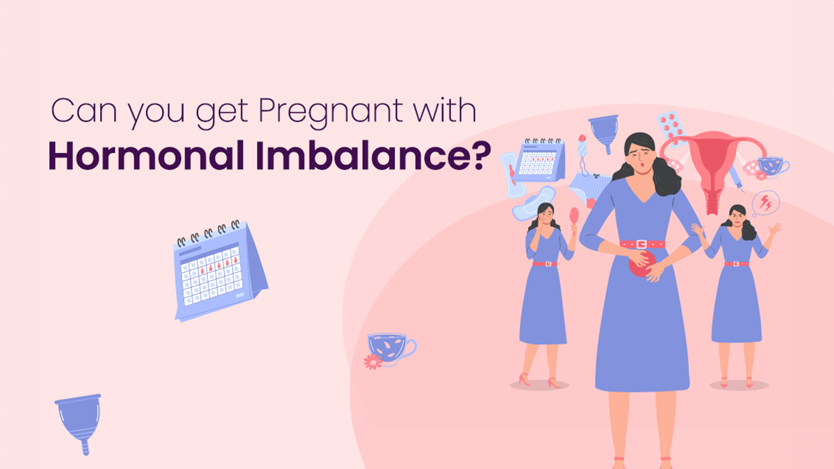
Are you struggling to conceive? Do you think hormonal imbalance might be the problem? Understanding how your body’s hormones affect your fertility is the first step to overcoming these challenges. Hormones control every part of reproduction. From your monthly cycles to pregnancy itself – hormones are in charge. An imbalance can disrupt this delicate system and make it harder for you to get pregnant. This article will explain how hormonal health and fertility are connected. It will help you identify symptoms, find effective treatments, and feel more confident on your journey to achieving pregnancy.
What is a Hormonal Imbalance?
A hormonal imbalance means that you have too much or too little of certain hormones in your bloodstream. Hormones serve as the body’s chemical messengers and hence, even small disruptions can have significant effects on how your body’s overall functions – especially when it comes to reproduction. Hormone imbalance in women can occur for various reasons. Factors such as your lifestyle, exposure to toxins, genetics, and medical conditions like polycystic ovary syndrome (PCOS) can all contribute.
Hormones Important for Pregnancy
When it comes to pregnancy, hormones like estrogen, progesterone, and testosterone play key roles. Estrogen regulates your monthly cycle and prepares your uterus for pregnancy. Progesterone supports the early stages of pregnancy. Even small amounts of testosterone are important for ovarian function.
How Hormonal Imbalance Affects Fertility
Hormonal imbalances create distinct fertility hurdles by disrupting important reproductive functions in multiple ways:
- Ovulatory Disruption:
- Ovulation becomes irregular or stops completely. Conditions like PCOS elevate androgens, preventing egg release.
- Egg quality suffers. Estrogen and progesterone imbalances lead to the development of nonviable eggs for fertilization.
- Irregularity of Menstrual Cycle:
- Periods arrive too frequently or infrequently. Fluctuating hormones shorten or lengthen cycles, affecting fertility windows.
- Menstruation halts completely. Severe imbalances cause amenorrhea (absence of menstruation), preventing natural conception.
- Endometrial Effects:
- Uterine lining fails to thicken adequately. Proper levels of estrogen and progesterone are necessary for building a nutrient-rich endometrial lining growth, crucial for implantation and supporting an early pregnancy.
- Defects in the luteal phase arise. Low progesterone levels shorten this post-ovulation phase, hindering the embryo’s ability to implant in the uterus.
Signs of Hormone Problems Hurting Baby Chances
Drastic weight fluctuations: When you gain or lose a lot of weight, it can impact hormones like estrogen and testosterone. These are important for maintaining reproductive health.
Too much hair growth or loss: These often indicate excess androgen levels, commonly seen in PCOS. Such high levels can affect ovulation and make it difficult to get pregnant.
Skin troubles: Acne and other skin issues might also signal hormone imbalance from excess androgens.
Feeling tired and moody: Though indirect, these could have a negative impact on the menstrual cycles.
Treatment Options for Hormonal Imbalance
1. Lifestyle Tweaks:
Addressing a pregnancy with hormonal imbalance often starts with lifestyle changes. Eating lots of fruits, vegetables, lean proteins, and whole grains, can positively impact the hormone levels. Exercise regularly to maintain a healthy weight since being over- or underweight can worsen hormone imbalances. Lastly, try managing your stress through yoga, meditation, and/or therapy as stress is known to throw off the hormone balance.
2. Medical Treatments:
Hormonal imbalances may require treatment beyond lifestyle adjustments. Treatment can include medicines for inducing ovulation in women with PCOS. Those facing thyroid hormone irregularities might get prescribed thyroid function regulators. Managing elevated prolactin levels could also involve specific hormonal imbalance drugs.
3. Assisted Reproductive Technologies (ART):
When natural conception is challenging due to hormonal imbalances, ART offers hope. Techniques like IVF and IUI can facilitate pregnancy by overcoming hormonal disorder barriers. However, fertility specialists should meticulously plan and execute such treatments, providing tailored advice based on individual hormonal health.
The Importance of Professional Guidance
Trying to conceive with hormonal imbalance requires professional insight. A hormone imbalance test, crucial for effective diagnosis and treatment, can be offered by healthcare providers. Working closely with fertility specialists or endocrinologists helps devise personalized treatment plans, including hormone imbalance therapy and other interventions tailored to specific patient needs.
Conclusion
Although a pregnancy with hormonal imbalance presents unique challenges, it is possible to conceive and have a healthy pregnancy with the right strategies and support. Women facing such difficulties are encouraged to seek guidance from healthcare professionals who are experts in hormonal health and fertility. By understanding the nature of hormonal imbalances and exploring all available treatment options, prospective parents can better their chances of achieving a successful pregnancy.


fill up the form to get a
Free Consultation
Avail 0% interest on EMI
All Procedures | No Upper Limit
Frequently Asked Questions
How can hormonal imbalance in women be curbed?
What are the symptoms of hormonal imbalance in women?
Can hormonal imbalance cause infertility?
What is the best time for hormone imbalance testing?
Related posts